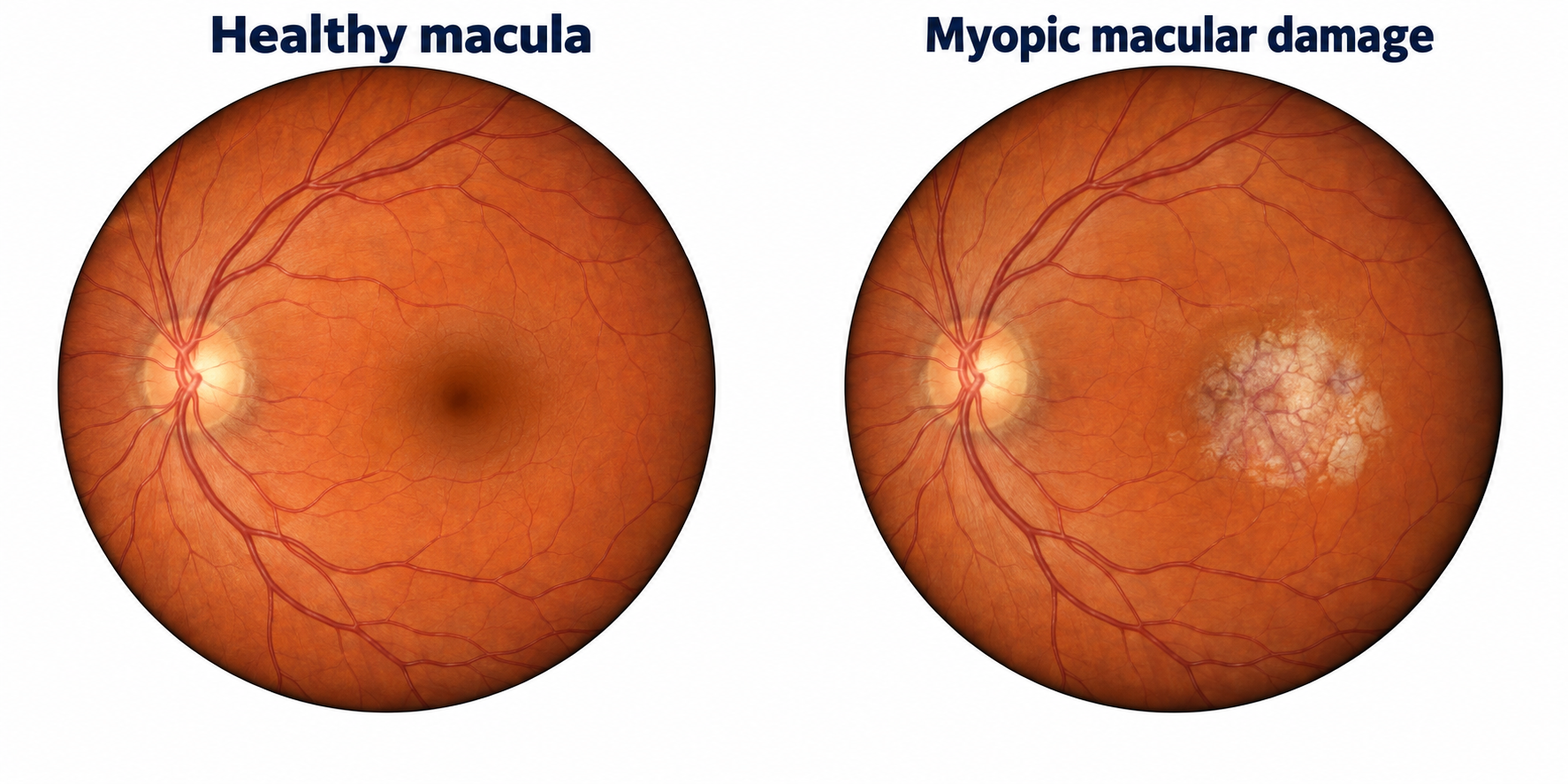
Myopic macular degeneration
The macula is the center of the retina. It helps us read and see faces. With severe myopia, this tissue and its support layers can thin and break down. Damage here can cause lasting loss of central sight. [6]
Eight practical sections to read, use, and bring to your child's next eye visit.
Download the parent guide (PDF)01 / NOTICE
A parent's guide to myopia and the sleepSEE Myopia Control system. Learn what changes to watch for, how care is planned, and how to track your child's progress.
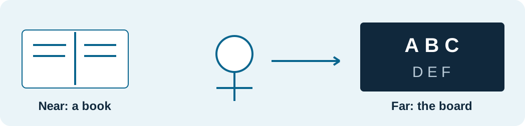
Your child may squint at the TV, miss words on the board, or say distant things look blurry. Tired eyes can be another clue. A child may not know that others see more clearly. Ask what feels hard to see. [1]
If your child needs stronger glasses again, bring the old and new prescriptions to the next visit. Ask what changed and how the doctor will track it. Do not guess how much the eyes have changed from symptoms alone.
Learn why eye growth matters (page 2). See retinal detachment (page 3) and other long-term risks (page 4). Prepare six questions (page 5). Get to know sleepSEE Myopia Control (page 6). Build a home routine (page 7). Keep a visit record (page 8).
Your next step: Write down one thing your child has noticed and when it began. Bring that note to a full eye exam.
02 / UNDERSTAND
Axial length is the eye's length from front to back. Eyes grow as children grow. With myopia, extra length can make distant things look blurry. That extra growth can also put future sight at risk. [3, 6]
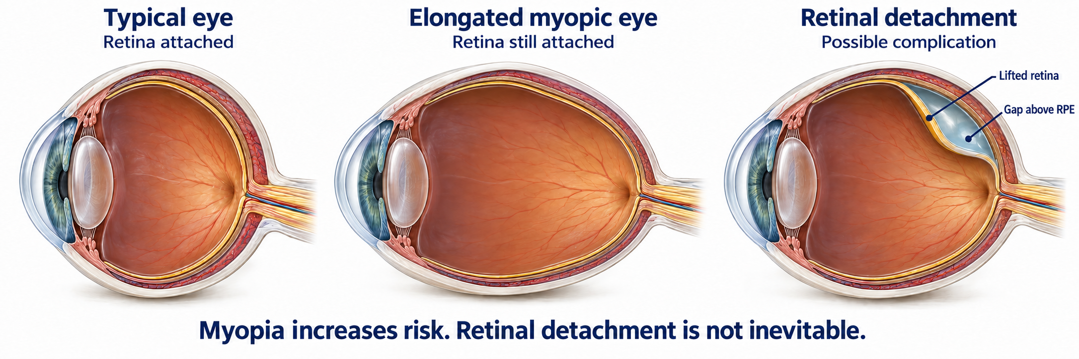
The retina is the light-sensing layer at the back of the eye. Parts of it can stretch and thin as the eye grows longer. These changes vary across the eye. They can make it more prone to damage. Clear vision with glasses or lenses does not prove that eye growth has slowed. [6, 8]
As myopia gets worse, the risk of these eye problems rises: [6, 7]
Myopia often gets worse faster when it starts early. It also has more years to get worse. Slowing extra eye growth early aims to limit how high myopia gets and lower later risks. Some eye damage can cause lasting sight loss. Stronger glasses cannot fix that damage. [3, 6]
These problems are possible, not certain. Your doctor can explain your child's risk. Ask: 'How much has each eye grown since our last visit, and does our plan need to change?'
Sources: [3] IMI: Clinical management guidelines · [6] IMI: Pathologic myopia and lasting vision loss · [7] National Eye Institute: Myopia and eye-health risks · [8] IMI: Eye-tissue changes with axial myopia · [9] AAO EyeWiki: Retinal detachment
03 / SEE THE DIFFERENCE
This close-up shows a retinal detachment. The retina has pulled away from its support layer. This is one possible problem linked to myopia. It does not happen in every eye with myopia. [6, 9]
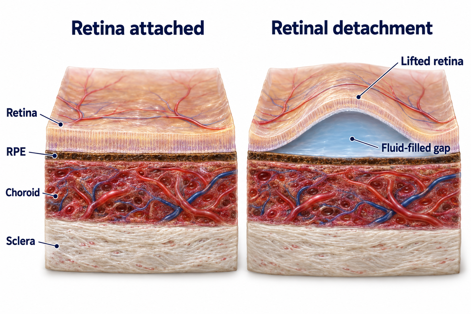
The RPE is a thin layer of support cells. The choroid below it carries blood that helps feed the outer retina. When the retina lifts away, that support is disrupted. Without prompt care, sight can be permanently lost. [9]
Higher myopia raises the risk that the retina may pull away. This is one reason to slow extra eye growth in childhood. The aim is to lower later risk. Care cannot promise that the retina will never pull away. [6, 7]
Get urgent eye care for sudden new flashes, many new floaters, or a curtain or shadow over vision. Do not wait for the next routine visit. [9]
Sources: [6] IMI: Pathologic myopia and lasting vision loss · [7] National Eye Institute: Myopia and eye-health risks · [9] AAO EyeWiki: Retinal detachment
04 / LOOK AHEAD
Myopia can raise the chance of these problems. They often start later in life. Higher myopia tends to mean higher risk. These problems are possible, not certain. [6, 7]
The macula is the center of the retina. It helps us read and see faces. With severe myopia, this tissue and its support layers can thin and break down. Damage here can cause lasting loss of central sight. [6]
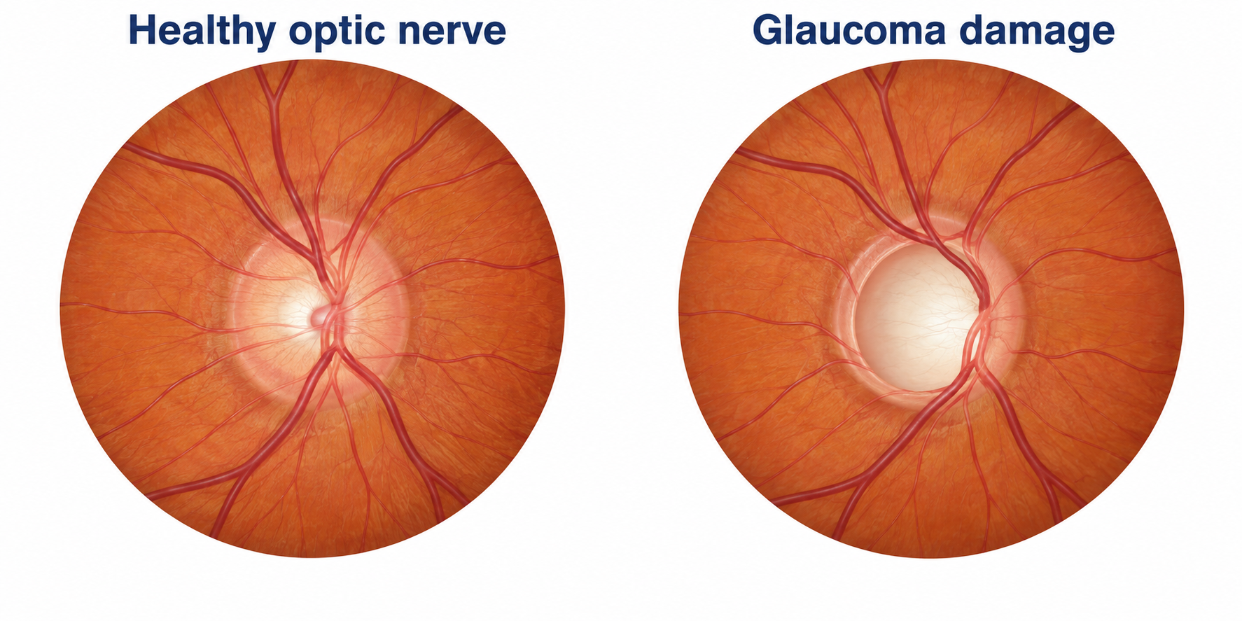
The optic nerve carries sight signals to the brain. Glaucoma damages this nerve and can slowly take away side vision. It often has no early symptoms. Regular eye exams can help find it before a person notices a change. [10]
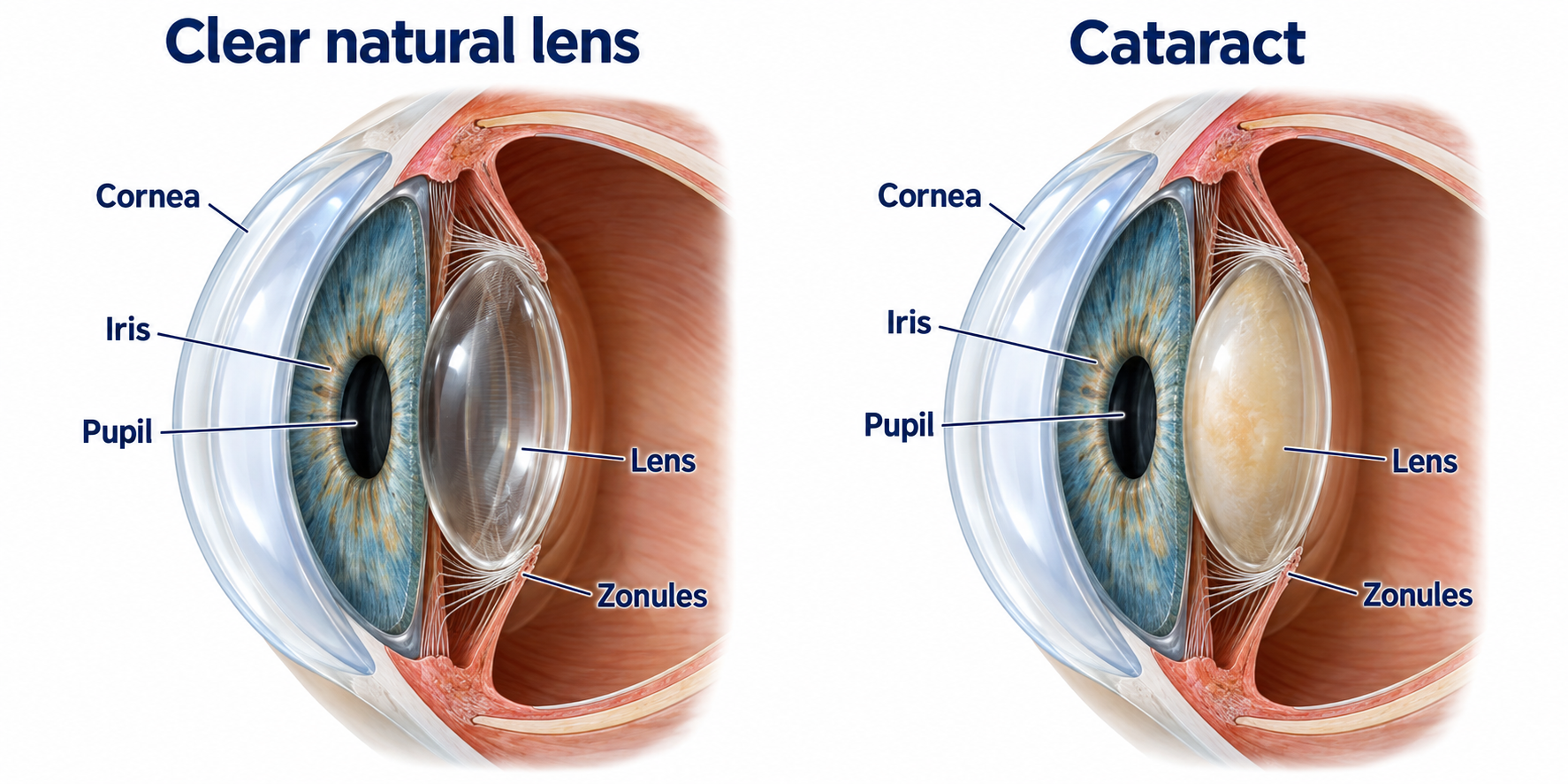
A cataract clouds the natural lens inside the eye. It can cause blur, glare, and trouble seeing at night. High myopia is linked to cataracts developing earlier. The cloudy part is the internal lens, not the cornea or a contact lens. [7, 11]
Illustrations show examples of tissue changes, not a prediction for your child. Details are enlarged for learning.
Some eyes grow new blood vessels under the retina. These can leak. Retinal layers may also split, or a hole may form in the macula. A bulge in the back wall of the eye is called a posterior staphyloma. This can affect sight. [6]
Ask your doctor how eye growth and eye-health checks guide your child's sleepSEE care plan.
Sources: [6] IMI: Pathologic myopia and lasting vision loss · [7] National Eye Institute: Myopia and eye-health risks · [10] National Eye Institute: Glaucoma · [11] National Eye Institute: Cataracts
05 / PREPARE
Bring past prescriptions, current glasses, and a list of concerns. Use the space below for short answers. Ask the doctor to explain any term you do not know.
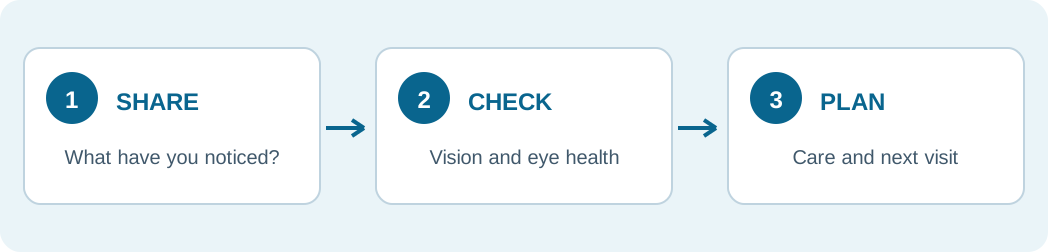
Before you leave: Ask for a clear plan, the next visit date, and the best number to call with questions.
06 / THE sleepSEE SYSTEM
sleepSEE Myopia Control is a system of care built around your child. It uses Ortho-K lenses as one part of a broader plan. Your doctor chooses the care steps and tracks how your child's eyes change.
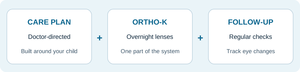
The doctor checks your child's vision, eye health, and eye shape. Bring past prescriptions and share your child's daily routine. Ask what their sleepSEE care plan will include and why.
Ortho-K lenses gently change the shape of the cornea, the clear front of the eye, during sleep. Your child takes them out in the morning. For children whose eyes fit the plan, Ortho-K can clear daytime blur and help slow myopia. [3, 4]
A prescription describes lens power. Repeat axial-length readings track eye growth. With Ortho-K, clear sight alone cannot show if growth has slowed. Corneal topography maps the eye's front surface. Your doctor uses all these tests to guide care. [2, 3]
The goal is to slow excess growth while keeping vision clear. It cannot promise to stop all growth or prevent every eye problem. Your doctor tracks each eye over time. Each visit helps show whether to keep or change the care plan. [3]
Follow the lens-care steps and home routine your doctor gives you. Keep each planned visit. Ask what is working, what has changed, and whether the care plan needs an update.
Lens care matters: Contact lenses can cause infection. Follow your doctor's care steps. If an eye is painful, red, or suddenly blurry, remove the lenses and contact the eye doctor promptly. [5]
Sources: [2] IMI: Myopia correction, control and management · [3] IMI: Clinical management guidelines · [4] IMI: Myopia-control treatments, 2025 · [5] FDA: Contact lens risks
07 / BUILD A ROUTINE
Use a routine your family can remember. sleepSEE suggests the schedule below for breaks from close work.
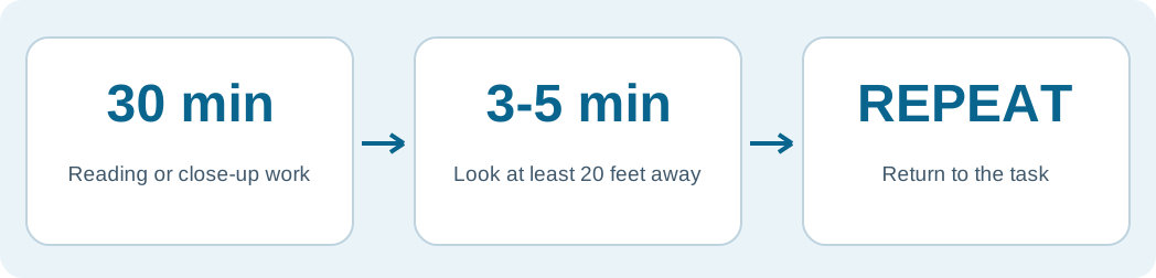
After 30 minutes of reading, homework, screen time, or other close-up work, have your child take a 3-5 minute break. During the break, have them look at something at least 20 feet away. Then they can return to the task and repeat the routine.
Set a timer. Choose a distant object before starting, such as a tree seen through a window. Put the book or device aside for the full break. A different close-up screen is not a distance break. Parents can use the same routine.
Make outdoor play part of the day when you can. More time outdoors can help lower the chance that myopia starts. Its role in slowing myopia after it starts is less clear. Breaks and outdoor time do not replace the care or eye checks your doctor directs. [3, 4]
For lens wear: Use the products and steps your eye doctor gives you. Keep contact lenses away from tap water, showers and swimming. Only sleep in lenses your doctor has prescribed for overnight wear. [3, 5]
Sources: [3] IMI: Clinical management guidelines · [4] IMI: Myopia-control treatments, 2025 · [5] FDA: Contact lens risks
08 / PRINT & KEEP
Print this page and bring it to each visit. Ask the care team to help fill it in. Use a new copy when you need more room. These are notes for discussion, not a way to diagnose changes yourself.
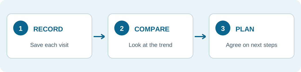
You can type in this worksheet, then print it. Entries stay on this page only; they are not sent or saved. Reloading clears them.
Ask about sleepSEE Myopia Control: Find a doctor in the program at sleepSEE.com/find-a-provider. Bring this guide and your child's old prescriptions. Discuss a plan for their eyes.