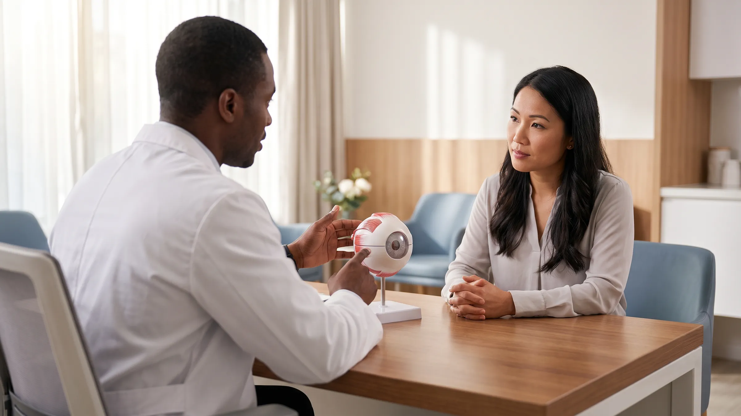

Axial length is the distance from the front to the back of the eye. Tracking it helps show how a child's eye is growing. It adds information to the prescription and eye health exam.
One reading is a starting point. The pattern over time helps the doctor assess myopia, or nearsightedness, and the response to care.
What is Axial Length, Really?
In myopia, light focuses in front of the retina, so distant things look blurry. The retina is the layer at the back of the eye that senses light. A myopic eye is often longer from front to back.
Eye length is measured in millimeters. Lens power also depends on the cornea and the lens inside the eye. The cornea is the clear front of the eye. There is no exact rule that links eye length to lens power for every person.
How We Measure It: A Look at Optical Biometry
An optical biometer uses light to measure the eye's length. The child looks at a target while the scan is taken. The test does not need to touch the eye.
Repeat readings help check that the test can be trusted. Ask how the practice takes readings the same way at each visit.
The Story of Growth: Normal vs. Concerning
Children's eyes grow, but the rate differs with age and other factors. A single annual-growth cutoff cannot decide every case.
The doctor may plot readings on a growth chart. This helps compare the trend with relevant data. The chart is one part of the exam, not a forecast of a child's final prescription.
| What is checked | What it adds |
|---|---|
| Axial length | Direct measure of front-to-back size |
| Prescription | The correction needed to focus clearly |
| Age and past readings | Context for the rate of change |
| Eye health and lens fit | Whether the current plan remains suitable |
Think of a child's height chart: one mark shows size today; several dated marks show growth. Eye-length records work in a similar way, though the doctor must use eye-growth data and the child's age. Two readings a month apart cannot be compared as if they were a year apart.
Ask to see the dates beside the numbers. Was the change steady? Were repeat scans needed to check a reading? What else changed in the exam? A small difference should be read with the test's limits in mind. IMI guidance on eye measurements.
The Risks of a Longer Eye: More Than Just Thick Glasses
Higher myopia is linked to greater risks of some eye diseases. These include:
- Retinal detachment: The retina pulls away from its normal place. New flashes, many new floaters or a curtain-like shadow need urgent eye care.
- Glaucoma: Damage to the nerve that carries signals from the eye to the brain.
- Myopic maculopathy: Damage in the central retina that can affect detailed vision.
- Cataract: Clouding of the lens inside the eye.
These are risks, not a prediction that one child will develop disease. A length measurement alone cannot give an exact lifetime risk. Keep eye health checks even when vision is clear.
Glaucoma can affect side vision and often has no early symptoms. That is one reason seeing clearly is not the same as having a full eye-health check. NEI: glaucoma.
Myopia can harm the center of the retina, the layer at the back of the eye. This damage can make it hard to read or see faces. High myopia does not always mean this damage is present. The doctor checks the eye for damage. The lens power alone cannot show it. IMI: pathologic myopia.
How sleepSEE Ortho-K Puts the Brakes on Growth
Ortho-K lenses change the cornea's shape during sleep. The child takes them out in the morning. The goal is clear daytime vision without daytime glasses or contacts.
The changed shape also changes light focus across the retina. This is thought to help signal slower growth. Research supports slower average eye growth in treated groups, but the response varies.
That is why your child's own measurements matter. Ask what has changed since the last visit and what would lead to a new plan.
| Stage | What to discuss |
|---|---|
| First exam | Baseline prescription, eye length and eye health |
| Follow-up | Repeat readings and daily vision needs |
| Plan review | Whether the trend supports continuing or changing care |
At the founding Fayetteville practice, the first-year Myopia Control / Management program includes eye-length checks and corneal maps twice a year. A corneal map shows the shape of the clear front of the eye. Your doctor sets the schedule for your eyes. See program inclusions and IMI care guidance. There are two separate checks: how clearly your child sees and how the eye grows. Ortho-K changes the front surface to improve focus. That means a clearer eye-chart result alone cannot tell you how much eye growth has slowed. Eye-length readings help answer the second question. Keep both parts of the record, even when your child is happy with daytime sight.
Dr. James Singletary, OD, FIAOMC
Dr. James Singletary is a licensed eye doctor. His main field is Ortho-K. He helps people see clearly without surgery. He also cares for children to help slow myopia, or nearsightedness. He created the sleepSEE Ortho-K program.
Medical Disclaimer
This article helps you learn about eye care. It is not personal medical advice, a diagnosis or a treatment plan. Ask a qualified eye doctor about your eyes and care. Do not use this page in place of that advice.